How to Recover From a Lumbar Disc Flare-Up
A lumbar disc flare-up can make everyday movement painful. Learn what may help, what can aggravate symptoms, when imaging is useful, and how an individualized plan supports recovery.

Meta description: Disc bulge vs herniated disc on MRI: learn what protrusion, extrusion, sequestration and nerve contact mean—and what actually matters for treatment.
If your MRI says disc bulge, protrusion, extrusion, or sequestration, it can sound like you have four completely different problems.
You don’t.
One of the most common conversations I have with patients is simply helping them understand what the words on their MRI actually mean.
A disc bulge and a herniated disc are not the same thing. Protrusion and extrusion are both types of disc herniation. Words like central, subarticular, and foraminal describe where the disc material is sitting. And words like contact, displacement, and compression describe what is happening near a nerve.
That may sound technical, but once you separate those ideas, an MRI report becomes much easier to understand.
More importantly, none of those words alone tells us how much pain you should have—or what treatment you need.
I often explain a spinal disc like an inner tube with thicker jelly in the middle.
The strong outer wall of the disc is called the annulus.
With a disc bulge, a broad portion of the disc pushes outward. In formal MRI terminology, a bulge generally involves more than half of the disc circumference.
Think of the wall of the inner tube widening or bowing outward over a larger area.
A herniated disc is different.
With a herniation, disc material moves beyond its normal boundary in a more localized area.
That distinction matters.
A bulge is not simply a “small herniation,” and a herniation is not simply a “bad bulge.” They describe different patterns of disc displacement.
You may also see the words “bulging disc” or “circumferential disc bulge” on an MRI report. Those generally describe that broader change in the disc contour.
Neither diagnosis automatically tells us how serious the problem is.
A disc bulge can hurt.
A herniated disc can sometimes cause very little pain.
What matters is where the finding is located, what structures it may be affecting, and whether it actually matches your symptoms and examination.
Here is another place MRI language can get confusing.
Radiologists sometimes describe how much of the edge of the disc is involved.
Focal generally means less than 25% of the disc circumference.
Broad-based generally means approximately 25% to 50%.
Think of that as describing how wide the abnormal area is around the edge of the disc.
The percentage helps describe the shape.
It does not tell us how much pain you should have, whether a nerve is involved, or whether you need surgery.
Those questions require the rest of the clinical picture.
This is where a lot of patients get lost.
Once we are talking about a herniated disc, two of the most common words you will see are protrusion and extrusion.
With a disc protrusion, the displaced material sticks outward, but the area where it remains attached to the main disc is generally wider than the portion sticking out.
Think of it as a localized bump that is still relatively broad where it connects to the disc.
With a disc extrusion, the displaced material extends farther beyond the normal disc boundary. The portion outside the disc may be wider than the area where it remains connected, or the material may extend in a way that otherwise meets the criteria for an extrusion.
The most important thing for a patient to remember is simple:
Protrusion and extrusion are both types of disc herniation.
An extrusion may look much more dramatic on an MRI, but seeing the word “extrusion” does not automatically mean you need surgery.
A 4 mm herniation also does not automatically hurt more than a 2 mm herniation.
Size matters, but location, inflammation, available space around the nerve, your symptoms, and your neurologic examination may matter even more.
Sometimes herniated disc material moves away from the exact spot where it originally came through the disc.
That is called migration.
The fragment can move upward or downward within the spinal canal and may still remain connected to the parent disc.
A sequestration goes one step further.
That means a piece of disc material has completely separated from the parent disc and become a free fragment.
Those words can sound alarming when you first read them on a report.
But remember what we are doing: we are describing anatomy.
We still have not answered whether that finding explains your symptoms or what treatment you need.
The annulus is the strong outer wall of the disc.
If some of those fibers separate, the MRI may describe an annular fissure.
You may hear people call this an annular tear, but “fissure” is generally the preferred imaging term.
An annular fissure can exist by itself or along with a disc herniation.
One important point: seeing a fissure on an MRI does not, by itself, tell us exactly when it occurred.
But that does not mean it is irrelevant.
The outer portion of the disc contains pain-sensitive nerve endings, and injured disc tissue can also create an inflammatory response.
That helps explain why sometimes a relatively small finding on MRI can still matter clinically.
Small on MRI does not always mean insignificant.
This is one of the biggest distinctions I explain when reviewing an MRI with a patient.
Words like bulge, protrusion, extrusion, and sequestration describe what the disc looks like.
Other words describe where it is sitting.
Common location terms include:
Central — toward the middle of the spinal canal
Paracentral or subarticular — just off center, near the lateral recess where a nerve root travels downward
Foraminal — in or near the opening where a nerve exits the spine
Extraforaminal — beyond that opening
Why does location matter?
Because nerves occupy very specific spaces.
A relatively small herniation sitting directly against a nerve may matter much more than a larger finding pointing into an area where there is plenty of room.
That is one reason we should never judge an MRI only by how many millimeters are written on the report.
This is another phrase that understandably gets patients worried.
An MRI report may describe the relationship between a disc and nerve with words such as:
Contact or abutment — the disc material is touching the nerve
Effacement — some of the normal space or fat around the nerve has been reduced
Displacement — the nerve has been pushed away from its normal position
Compression — the nerve appears clearly pressed or flattened
Those are not all the same thing.
“Contacting a nerve” is not the same as severely compressing it.
And even when there is true nerve compression, we still have to ask what is happening to the patient.
Do you have pain traveling into the leg or arm?
Is there numbness or tingling?
Is strength changing?
Are reflexes abnormal?
Does the side and level of the MRI finding actually match those symptoms?
That is when the picture on the MRI starts becoming clinically useful.
Here is a realistic lumbar MRI finding:
“L4-L5 left subarticular disc extrusion contacting the traversing left L5 nerve root.”
That sounds intimidating until we translate it.
This is the disc between the fourth and fifth lumbar vertebrae.
The herniation is sitting just off center on the left, in the lateral recess where a nerve root travels downward.
The shape of the herniation meets the criteria for an extrusion.
The herniated material is touching the L5 nerve root as that nerve travels downward toward where it eventually exits the spine.
Now the sentence makes much more sense.
It tells us what the disc looks like, where it is located, and which nerve is nearby.
What it does not tell us by itself is:
how much pain you should have
whether the disc explains every symptom
exactly what caused the finding
when it occurred
whether it will improve
whether you need surgery
whether another structure is contributing to your pain
Those questions require more than the MRI report.
They require the patient.
Not by itself.
MRI is excellent at showing anatomy.
But an MRI is still a picture.
Two people can have remarkably similar MRI findings and completely different symptoms.
One person may have significant leg pain, numbness, or weakness.
Another person may barely know the herniation is there.
The opposite can happen too. Someone can have significant pain even when routine imaging does not look particularly dramatic. I explain that concept further in why clear X-rays can still miss mechanical spine pain.
The goal is not simply to find something abnormal on imaging.
The goal is to determine whether the abnormality explains what is happening to you.
No.
The terminology on the MRI helps us understand the anatomy, but treatment decisions depend on much more.
We look at things such as:
where your symptoms travel
strength
sensation
reflexes
walking and daily function
how your spine responds to movement and load
the level and location of the disc finding
whether the MRI and neurologic examination actually match
Many disc problems can initially be managed conservatively when neurologic function is stable and emergency findings are absent.
Other cases need pain management, orthopedic spine evaluation, neurosurgery, or additional testing.
And sometimes surgery is exactly the right treatment.
The goal should not be to avoid surgery at all costs.
The goal is to determine which level of care gives that individual patient the best path forward.
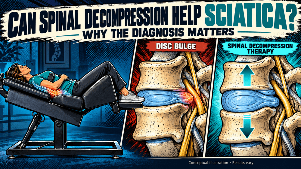
For selected patients with disc-related pain, spinal decompression therapy in Charleston may be one conservative option. Candidate selection matters, which is why we first need to understand the MRI, neurologic findings, symptoms, and overall clinical picture.
If you are already weighing those decisions, I have also explained spinal decompression vs. surgery for a herniated disc in more detail.
This is exactly why I believe treating an MRI report by itself is a mistake.
At Elite Family Chiropractic, we evaluate the disc finding in the context of the entire case.
Where is the herniation?
Which nerve is nearby?
Does it match your pain pattern?
Is strength changing?
Are reflexes or sensation abnormal?
How is the spine tolerating movement and load?
Could another structure be contributing to the pain?
From there, we can determine whether an individualized conservative treatment plan makes sense or whether another level of care is more appropriate.
For some patients, that may include chiropractic care, rehabilitation strategies, activity modification, or spinal decompression when appropriate.
For others, the most important thing we can do is recognize that the case needs collaboration with physical therapy, pain management, orthopedic spine, neurosurgery, or another provider.
That is how I believe complex spine care should work.
Understand the problem first. Then choose the treatment.
No. A disc bulge describes a broader outward change in the disc contour. A herniated disc describes more localized displacement of disc material beyond its normal boundary.
Yes. Protrusion and extrusion are two different shapes of disc herniation.
It can represent a more substantial structural finding, but the word “extrusion” alone does not tell you how severe your symptoms should be or whether surgery is necessary. Location, nerve involvement, neurologic findings, and function matter.
Subarticular describes the location of the herniation. It sits off center in the lateral recess, an area where a traversing nerve root may travel.
Not necessarily. Contact means the disc material is touching the nerve. Displacement or compression describes a greater degree of mechanical effect.
Yes. MRI findings and symptoms do not always match perfectly. That is why the MRI should be interpreted alongside your symptoms and examination.
If your report says disc bulge, protrusion, extrusion, sequestration, subarticular, foraminal, or nerve root contact, do not judge the entire problem from one word.
First understand:
What shape is the disc finding?
Where is it located?
What is it doing near the nerves?
Then ask the most important question:
Does that anatomy actually match what is happening to me?
That is where an MRI report stops being a collection of confusing medical terms and starts becoming useful information for deciding what to do next.
If you have been diagnosed with a disc bulge or herniated disc and are not sure what your MRI means for you, learn more about our approach to herniated disc care in Charleston.
At Elite Family Chiropractic, our goal is to help you understand the problem, build an individualized plan, and make sure you get the level of care you need—whether that means conservative treatment with us or collaboration with another spine specialist.